2026 Comprehensive Parent Guide
As a parent, we perfectly understand the protective instinct you feel when your child sits in the dental chair. When your dentist turns and says, “We need to take a small x-ray before starting treatment,” that sudden anxiety is one of the most common feelings for modern parents.
“My child is still very young, is it right for them to receive this much radiation?”, “Can’t you just check visually?”, “Will these rays harm their brain or thyroid gland?” It is perfectly natural for such questions to cross your mind.
At Prof. Dr. Behiye Bolgül Clinic, transparency is our most important principle. In this comprehensive guide, we will set aside urban legends and focus solely on scientific data, standards from international health authorities (WHO, ADA, ICRP), and the facts about digital radiography.
Related Articles
- What If a Primary Tooth Is Lost Early? Space Maintainer Guide
- Dental Abscess in Children: Symptoms and Treatment
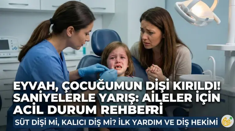
- Dental Trauma Treatments in Children
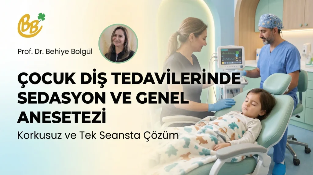
- Sedation and General Anesthesia in Pediatric Dentistry
- Baby Bottle Tooth Decay: Causes and Prevention
1. The Invisible Side of the Iceberg – Why Must We Take X-Rays?
Many parents think dentistry is only about “visible teeth”. However, dentistry is a field where millimeter details are vital, and a clinical examination (visual check) only allows us to see the tip of the iceberg.
A diagnosis made without x-rays is like driving a car blindfolded.
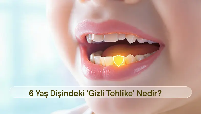
The Hidden Enemy: Interproximal Decay
Children’s tooth enamel is thinner and more permeable than adults’. Decay starting at the contact points between two teeth may look pristine from the outside. However, on an x-ray, it is clearly visible that the tooth is being hollowed out from the inside. If an x-ray is not taken, this tooth will suddenly manifest with severe pain (pulpitis) one night.
Root Development and Infections
Even if the tooth color doesn’t change after a fall or impact, there may be fractures in the root. Or a deep cavity may have formed a silent abscess within the jawbone. We can only see these with an x-ray.
Developmental Monitoring and Missing Teeth
Some children may have congenitally missing teeth (agenesis) or extra teeth (supernumerary). The position of permanent teeth under the baby teeth signals the need for orthodontic treatment years in advance.
2. Radiation Phobia and Scientific Facts
The biggest source of fear is the word “radiation”. However, radiation is a natural part of our lives. We receive natural radiation every day from the sun, soil, buildings, and even the food we eat. This is called “Background Radiation”.
To understand the dose your child receives from a dental x-ray, let’s compare it with other daily activities.
| Activity / Situation | Approximate Radiation Dose (μSv) |
|---|---|
| Eating 1 Banana | 0.1 μSv |
| Digital Dental X-Ray (Single Tooth) | 0.1 – 0.5 μSv |
| Daily Natural Background Radiation | 8 – 10 μSv |
| Flight (Istanbul – New York) | 40 – 50 μSv |
| Chest X-Ray (Hospital) | 20 – 40 μSv |
| Panoramic Dental X-Ray (Full Jaw) | 10 – 25 μSv |
Striking Fact: The single small digital dental x-ray your child has at the dentist is equivalent to the natural radiation they receive playing in the park for 1-2 hours on a sunny day. So, refusing treatment to protect your child from a dental film is as extreme as never taking them outside to protect them from the sun.
3. Technology Has Changed: The Shift from Analog to Digital
In the past (analog era), films had to be developed and exposure times were long. However, modern pediatric dental centers now use Digital Radiography (RVG) technology.
- 90% Less Radiation: Digital sensors are much more sensitive to X-rays. This reduces the required radiation amount by up to 90%.
- Image in Seconds: No waiting for film development. The image appears on the screen in seconds.
- Image Processing: Your dentist can magnify the image, adjust contrast, and examine details without needing to retake the x-ray.
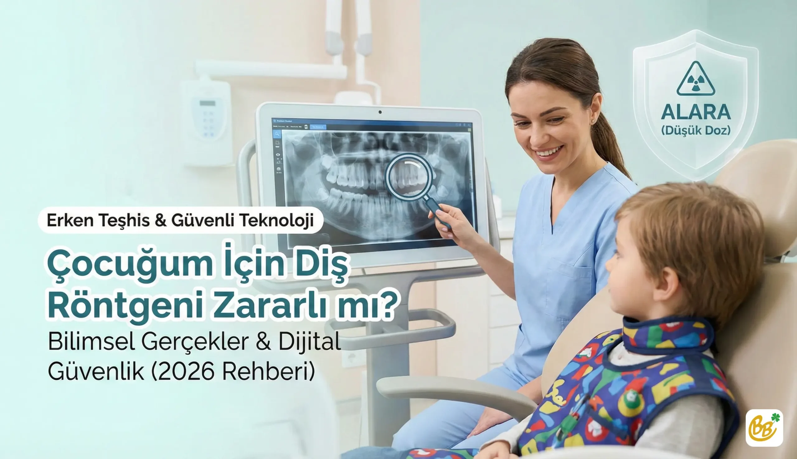
4. Safety Protocol for Children: The ALARA Principle
Dentists worldwide operate according to the ALARA (As Low As Reasonably Achievable) principle.
- Only If Necessary: X-rays are not taken routinely for everyone, but only when suspected during a clinical examination.
- Lead Apron and Thyroid Shield: During the scan, your child is always fitted with a special lead-lined apron and collar. This protects the rest of the body.
- Fast Sensors: Children move. Fast digital sensors prevent film blurring due to movement and the need for retakes.
- Child Mode: The “pediatric mode” found on devices automatically lowers the radiation dose according to the child’s bone structure.
5. Which X-Ray is Taken for What?
There are different types of x-rays depending on your child’s needs:
- Periapical (Single Tooth) X-Ray: Shows 2-3 teeth and their roots in detail. Used for deep decay, root canal treatments, and trauma.
- Bitewing X-Ray: The best and lowest dose method for detecting “hidden interproximal decay”.
- Panoramic X-Ray: Shows the entire jaw structure, baby teeth, erupting permanent teeth, and impacted teeth in a single film. Usually taken for a general check-up after age 6.
Risk Analysis: To Take or Not to Take?
Let’s explain with an example:
Scenario A (X-Ray Taken): The dentist caught the invisible early decay between two teeth. Treatment was completed in 15 minutes with a preventive filling, without even needing anesthesia. The tooth remained vital.
Scenario B (X-Ray Refused): The decay was not noticed. 6 months later, the child woke up with severe pain. A dental abscess formed, antibiotics were used, and perhaps the tooth had to be extracted.
Result: The harm caused by an untreated dental infection to your child is thousands of times greater than the theoretical radiation risk of a digital x-ray.
Frequently Asked Questions
Baby teeth will fall out anyway, is it worth taking an x-ray?
Absolutely. An infection in a baby tooth can damage the enamel of the permanent tooth developing underneath. Early extraction of a baby tooth leads to space loss and future orthodontic problems. Early diagnosis saves lives (and teeth).
My child has a gag reflex, how do you take films?
Pediatric dentists are trained in this. Special sized sensors are used for small mouths. If necessary, extraoral methods are preferred.
How many times a year can x-rays be taken?
There is no exact number, it depends on the child’s “caries risk group”. High-risk children may need a check-up film every 6 months, while for low-risk groups, once every 1-2 years is sufficient.
Knowledge Conquers Fear
“Instead of ‘guessing’ what lies beneath your child’s beautiful smile, we safely use x-ray technology to ‘be sure’ and offer the most protective treatment. Remember, early diagnosis not only saves teeth; it also improves your child’s experience in the dental chair.”— Prof. Dr. Behiye Bolgül